
Doctors in China inadvertently took time-series images of parasitic worms actively invading a woman's brain and causing rare and rapidly progressing lesions.
The previously healthy 60-year-old woman went to the hospital after having a fever and altered mental status for three days, according to a report of her case published Monday in JAMA Neurology. By the time she arrived, she was unable to communicate normally.

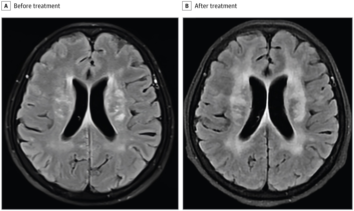
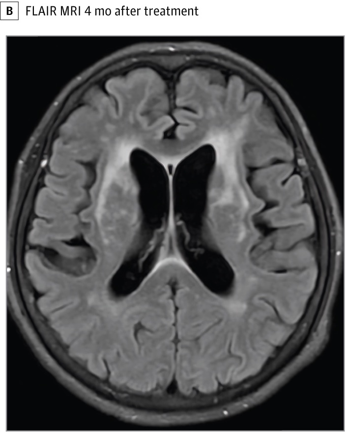
Magnetic resonance imaging (MRI) showed white matter lesions around her lateral ventricles, large cavities in the center of the brain filled with cerebrospinal fluid that, from the side, are C-shaped. The type of MRI used, a FLAIR MRI, is used to more easily detect lesions, and the fluid-filled lateral ventricles appear as dark, curved spaces in the center. Doctors could see white blotches and smears around those dark spaces, indicating lesions. After doing a spinal tap and running tests on her cerebral spinal fluid, they suspected she might have a bacterial infection in her brain. So they treated her with an antibiotic and a fever reducer.
But two weeks later, her symptoms hadn't improved. The doctors repeated a FLAIR MRI and found that the white lesions had only gotten worse. They did more tests.
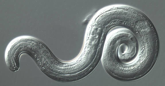
Blood tests showed elevated eosinophils, which can be a sign of a parasitic infection, among other things. They searched for genetic signatures of parasites in her cerebrospinal fluid and got a strong hit. The genetic sequencing matched Angiostrongylus cantonensis, also known as rat lungworm.
Sickening cycle
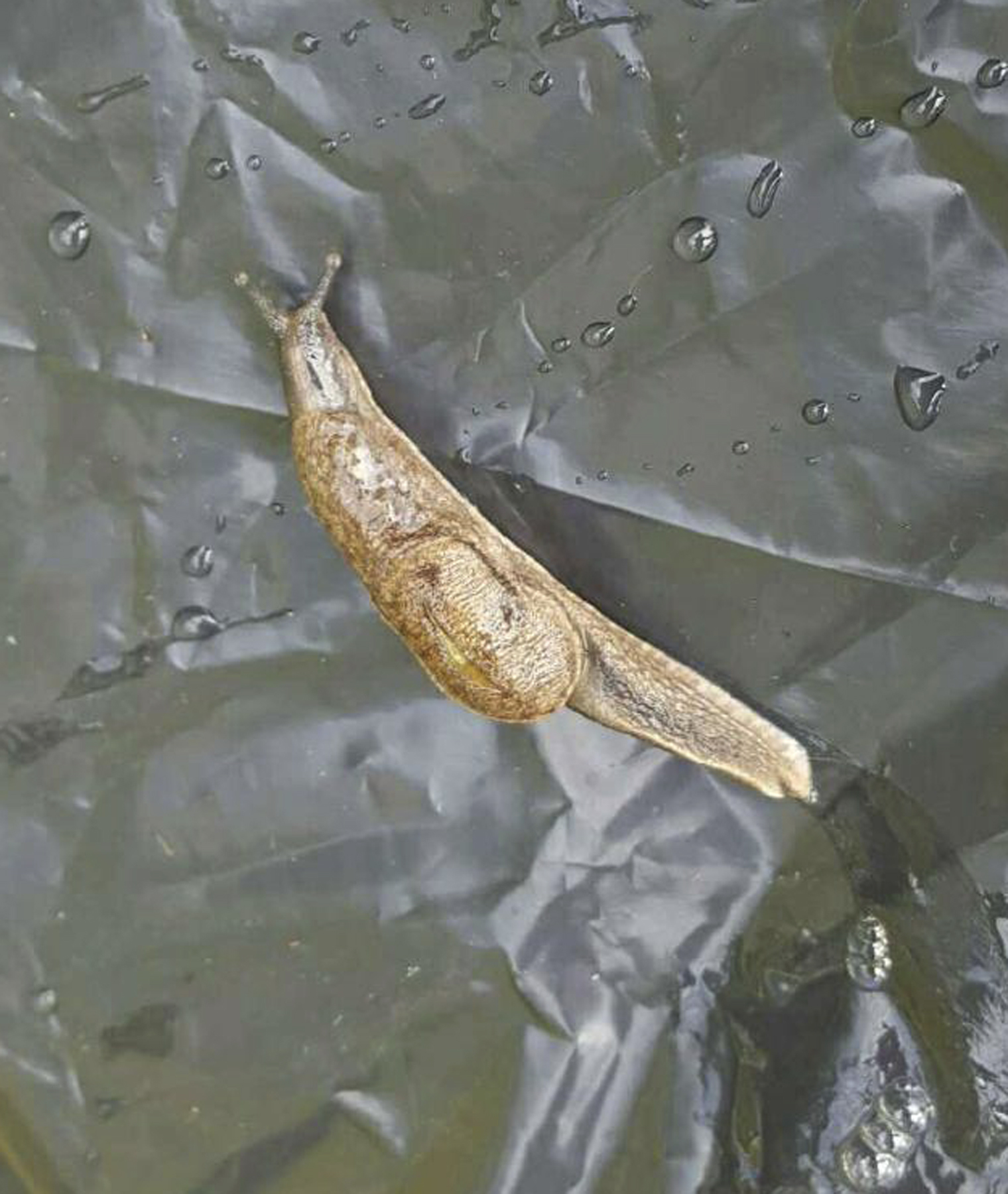
This ghastly parasite causes outbreaks and sporadic cases in various places around the world, including China and other parts of Asia. You may remember hearing of it amid an uptick in cases reported in Hawaii a few years ago or a report in 2023 that it had invaded the Southeast US.

 Loading comments...